Implants
Periodontics
More
805-312-8604

Advanced Care for Your Gums & Implants

Specializing in periodontal disease treatment, dental implants, and gum recession — delivering precision care with compassion. Proudly serving Camarillo, Oxnard, Ventura, and surrounding areas.

Modern dental office

Comprehensive Periodontal & Implant Services

From routine gum disease treatment to full-arch implant restorations, we offer the full spectrum of periodontal specialty care.

Dental Implants

Gum Grafting

Full Arch (All-on-X) Implants

Bone Grafting

Flexible Payment Options

Treatment Financing Options

We work with leading financing partners to make your care affordable.

CareCredit Cherry LendingClub Proceed Finance

Ready to Get Started?

Your Healthy Smile Begins Here

Schedule a consultation and take the first step toward exceptional periodontal health.

Home › Meet the Doctor

Dr. Ramin Foroughi

Periodontist · Limitless Periodontics & Implants

Dr. Ramin is a periodontist dedicated to combining advanced technology with compassionate care. He is trained in some of the most modern techniques in dentistry, including robot-guided and fully guided implant surgery and microsurgery, allowing him to deliver highly precise and predictable results.

He completed his undergraduate degree from UCLA in 2013 and his dental degree at the University of Southern California in 2017. He practiced as a general dentist for four years before completing his Periodontal specialty at Boston University — where he also taught Periodontics to dental students — giving him a strong restorative foundation that enhances his surgical approach and helps ensure the best esthetic outcomes for patients.

Beyond his clinical expertise, Dr. Ramin has volunteered in multiple Dental Humanitarian Outreach Programs (DHOP) through USC in the Philippines, Cambodia, Panama, and Belize, treating patients in some of the most underserved areas of the world. He was directly involved in organizing several of these trips and served as faculty teaching USC dental students.

Dr. Ramin Foroughi at a DHOP humanitarian mission

Dr. Ramin treating a patient during a DHOP humanitarian mission abroad.

Dr. Ramin prides himself on clear and open communication, making sure every patient feels as comfortable and informed as possible throughout their care. Outside of dentistry, he enjoys working out and hosting dinners with friends.

Education & Training

  • B.A., University of California, Los Angeles (UCLA) — 2013
  • D.D.S., University of Southern California (USC) — 2017
  • Certificate in Periodontics, Boston University — Advanced Specialty Education
  • Training in Robot-Guided & Fully Guided Implant Surgery
  • Advanced Training in Microsurgery

Professional Memberships & Service

  • American Dental Association (ADA)
  • California Dental Association (CDA)
  • Santa Barbara-Ventura County Dental Society
  • Dental Humanitarian Outreach Program (DHOP) — Philippines, Cambodia, Panama & Belize

Member Organizations

American Dental Association California Dental Association Santa Barbara-Ventura County Dental Society
Periodontist

Specialty Matters

Why a Periodontist?

A periodontal residency is a rigorous, full-time three-year program dedicated entirely to the surgical procedures we perform every day — dental implants, gum surgery, bone grafting, and the structures that support your teeth. During those three years, that is all we do. We are not learning a little bit of everything; we are mastering a very specific set of skills with thousands of hours of hands-on training under expert supervision.

By contrast, many general dentists who perform implants and gum procedures have attended weekend courses — two or three days of instruction before returning to practice. We respect our general dentist colleagues deeply, and many are talented clinicians. But a weekend course cannot replicate years of focused surgical training.

The data reflects this difference. Studies consistently show that implants placed by specialists have lower failure rates and fewer long-term complications than those placed without equivalent training. Many implants placed with limited experience may appear fine in the short term — but they are significantly more likely to fail over time. When it comes to a procedure that is meant to last a lifetime, experience and specialization are not just a preference. They are the standard of care.

Home › Implants › Extraction & Socket Preservation
The Condition

When a Tooth Cannot Be Saved

Sometimes a tooth is too damaged, decayed, or infected to be restored. In these cases, extraction is the right decision — but what happens immediately after the tooth is removed is just as important as the extraction itself.

When a tooth is lost, the surrounding bone begins to resorb within days. Without the tooth root stimulating the jawbone, up to 50% of bone volume can be lost within the first year. This bone loss can complicate or even prevent future implant placement, requiring more extensive and costly procedures down the road.

Acting at the time of extraction gives us the best opportunity to preserve the bone that is already there — setting the foundation for a seamless implant in the months ahead.

Socket preservation procedure
Our Approach

Extraction & Socket Preservation

Socket preservation (also called alveolar ridge preservation) is a procedure performed at the time of tooth removal. Bone grafting material is placed directly into the empty socket immediately after extraction to maintain the shape and volume of the jawbone while it heals.

  • 1

    Gentle Extraction

    The tooth is carefully removed with techniques that minimize trauma to the surrounding bone and soft tissue.

  • 2

    Socket Grafting

    Bone graft material is placed into the socket immediately, filling the space left by the tooth root and preventing collapse.

  • 3

    Membrane Placement

    A protective collagen membrane is placed over the graft to shield it and guide proper bone regeneration.

  • 4

    Healing Period

    The site heals over approximately 4 months, allowing the graft to mature into solid, healthy bone ready to support an implant.

Socket preservation bone grafting
Why It Matters

Benefits of Socket Preservation

  • Prevents significant bone loss after extraction
  • Maintains ideal ridge shape for future implant placement
  • Reduces the need for more extensive bone grafting later
  • Preserves the natural contour of your gum and jaw
  • Performed at the same appointment as the extraction
  • Minimizes overall treatment time and cost
  • Creates the best possible foundation for a life-long implant
Patient after socket preservation
Home › Implants › What Are Dental Implants?
The Condition

Living With Missing Teeth

When a tooth is lost — whether from decay, gum disease, or injury — the consequences go far beyond appearance. The surrounding bone begins to shrink immediately, neighboring teeth drift, and bite force drops significantly. Over time, even facial structure can change.

Traditional bridges require grinding down healthy adjacent teeth. Removable dentures slip, limit diet, and continue to allow bone loss underneath. Neither solution addresses the root cause: the absence of a tooth root stimulating the jaw.

Without intervention, bone loss accelerates each year — making future treatment more complex and costly. The longer the wait, the more bone is lost.

Dental implant cross-section showing crown, abutment, titanium post and jawbone
Our Solution

What Is a Dental Implant?

A dental implant is a small titanium post that replaces the missing tooth root — permanently anchored into the jawbone. It fuses with bone through osseointegration, creating a stable, lifelong foundation for a crown, bridge, or denture.

  • 1

    Titanium Post (Implant Fixture)

    Surgically placed into the jawbone exactly where the tooth root once was.

  • 2

    Abutment Connector

    Attaches to the top of the implant post once osseointegration is complete.

  • 3

    Custom Porcelain Crown

    Precision-matched to your natural teeth — indistinguishable in look and function.

3-part dental implant diagram showing Crown, Abutment, and Implant post
Why It Matters

Benefits of Dental Implants

  • Look, feel, and function like natural teeth
  • Preserve jawbone and prevent facial collapse
  • No need to alter adjacent healthy teeth
  • Restore full chewing force
  • Eliminate embarrassing slippage
  • Brush and floss normally — no special tools
  • 95%+ long-term success rate
Patient smiling after dental implant treatment
Home › Implants › Dental Implant Placement
The Need

When Is Implant Placement Needed?

Tooth loss — from decay, fracture, gum disease, or trauma — leaves a gap that affects your bite, speech, and confidence. More critically, the underlying bone begins resorbing within weeks of extraction, making early placement essential.

Unlike a bridge that spans the gap or a denture that rests on the gum, an implant directly replaces the missing root. This is the only solution that stops bone loss.

Early intervention preserves maximum bone volume, simplifies the surgical procedure, and provides the best long-term outcomes. The sooner an implant is placed, the easier the process.

Dental implant placement cross-section
The Procedure

Implant Surgery — Step by Step

Using 3D CBCT imaging and digital surgical guides, Dr. Ramin custom plans every implant before the first incision is made. When a tooth still needs to be removed, the full process from extraction to final crown can take up to 9 months — and possibly longer depending on individual healing.

  • 1

    3D Imaging & Planning

    CBCT scan maps bone volume, density, and anatomy for precise digital planning and to assess whether extraction and bone grafting are needed before implant placement.

  • 2

    Tooth Extraction (when needed)

    If a failing or damaged tooth is still present, it is gently removed. Whenever possible, a socket preservation bone graft is placed at the same appointment to maintain the bone volume needed for the implant.

  • 3

    Bone Graft Healing — ~4 Months

    After extraction and grafting, the site is allowed to heal for approximately 4 months. During this time the graft matures into solid bone capable of supporting an implant. A temporary solution may be provided during this phase.

  • 4

    Anesthesia & Comfort

    Local anesthesia and optional sedation ensure a completely pain-free implant surgery experience.

  • 5

    Implant Placement

    Dr. Ramin follows the plan of his 3D planning to direct the drill to the planned position and angle. The titanium implant is threaded in with calibrated torque for immediate stability.

  • 6

    Osseointegration (3–6 months)

    The implant fuses with the surrounding bone, forming a permanent root-like anchor. In cases requiring extraction and grafting, the total timeline from start to crown can reach 9 months or longer.

  • 7

    Crown Delivery

    Once osseointegration is confirmed, your restorative dentist attaches a custom crown. Your new permanent tooth is complete.

Implant anatomy diagram
Why Choose a Periodontist

Benefits of Specialist-Placed Implants

  • Specialist training in bone and gum anatomy
  • Lower complication and failure rates
  • Bone grafting expertise when needed
  • In-office sedation available
  • Immediate loading protocols when eligible
  • Predictable, long-term results
  • Highest standard of surgical precision
Patient smiling after implant placement
Home › Implants › Sinus Lift
The Condition

Not Enough Bone for Upper Jaw Implants?

When back teeth in the upper jaw are lost, the surrounding bone resorbs and the maxillary sinus — the large air cavity just above — gradually expands downward into that space. This process, called sinus pneumatization, can leave as little as 3–4 mm of bone where a minimum of 8mm is needed for an implant.

Many patients in this situation have been told they cannot have implants in the upper back jaw. A sinus lift (sinus augmentation) changes that — rebuilding the bone platform needed for safe, lasting implant placement.

The good news: approximately 90% of patients who need a sinus lift only need the simpler internal approach, which can be performed at the same time as implant placement — with no additional healing time and no noticeable difference in the patient experience.

Internal sinus lift with simultaneous implant placement
~90% of Patients
The Procedure

Internal Sinus Lift

Performed entirely through the implant site itself using a gentle osteotome technique. The implant is placed at the exact same appointment — patients typically notice no difference compared to a regular implant procedure.

  • 1

    3D CBCT Imaging

    Precisely measures residual bone height and sinus anatomy to confirm the internal approach is appropriate.

  • 2

    Osteotome Elevation

    Through the implant socket, a specialized instrument gently lifts the sinus floor just enough to create space for the bone graft and implant.

  • 3

    Bone Graft Placement

    A small volume of bone graft material is placed to fill the newly created space beneath the sinus membrane.

  • 4

    Same-Day Implant Placement

    The titanium implant is immediately inserted at the same appointment. No additional healing phase is required before placement.

  • 5

    Osseointegration & Crown

    The implant integrates with surrounding bone over 3–6 months, then your restorative dentist places the permanent crown.

Internal sinus lift procedure
~10% of Patients
The Procedure

Lateral Sinus Lift

Reserved for cases with very minimal residual bone (under 4 mm). A small window is carefully made in the side wall of the sinus, allowing a larger volume of bone graft to be precisely placed. This approach requires a separate healing phase before implants can be placed.

  • 1

    3D CBCT Imaging

    Confirms that residual bone is under 4 mm, requiring the lateral window approach.

  • 2

    Lateral Window Creation

    A small access window is created in the lateral wall of the sinus under local anesthesia, allowing direct visualization and access to the sinus cavity.

  • 3

    Large-Volume Bone Graft

    The sinus membrane is elevated and a substantial volume of bone graft is packed into the space. A collagen membrane covers the window to protect the graft.

  • 4

    Graft Maturation (4–9 Months)

    The site heals and the graft matures into mature bone volume sufficient for implant anchorage.

  • 5

    Implant Placement & Crown

    Once sufficient bone volume is confirmed by CBCT, implants are placed and the restoration proceeds as normal.

Lateral sinus lift surgical procedure
Why It Matters

Benefits of a Sinus Lift

  • 90% of patients treated with a single same-day procedure
  • Implant placed at the same visit in most cases
  • Enables implants where previously impossible
  • Long-term bone volume preservation
  • 95%+ predictable success rate
  • Eliminates need for removable partial dentures
Patient smiling after sinus lift and implants
Home › Implants › Implant-Supported Overdenture
The Condition

The Struggles of Traditional Dentures

Conventional removable dentures rest on the gum ridge and are held in place by suction or adhesive paste. While they restore appearance, they fall dramatically short on function. Studies show that denture wearers retain only 20–30% of their original chewing force, making it difficult — sometimes impossible — to eat fibrous vegetables, meats, or crusty breads. This dietary restriction carries real nutritional consequences over time.

Beyond eating limitations, loose dentures create social anxiety. Slippage while speaking, laughing, or eating in public is a common source of embarrassment that leads many patients to withdraw from social situations. Repeated movement also causes chronic sore spots and denture stomatitis from friction against the gum tissue.

Perhaps most critically, traditional dentures do nothing to address ongoing bone loss. Without tooth roots or implants stimulating the jawbone, the ridge beneath the denture continues to shrink — often requiring new dentures every few years as fit deteriorates. An implant-supported overdenture solves all of these problems at a far more accessible price point than a fully fixed restoration.

Implant-supported overdenture snap-in attachment
The Procedure

How We Treat It

An implant-supported overdenture snaps onto 2–4 strategically placed implants using precision ball attachments or a rigid locator bar. The denture is removable for easy cleaning, yet locked firmly in place during use — eliminating all movement and restoring genuine confidence.

  • 1

    Comprehensive Planning

    CBCT 3D imaging evaluates bone volume and density to determine the optimal number and position of implants for your jaw anatomy.

  • 2

    Implant Placement

    Two to four titanium implants are placed at precisely planned positions in the jawbone under local anesthesia and optional sedation.

  • 3

    Osseointegration Healing

    Over 3–6 months the implants fuse securely with the surrounding bone, establishing the stable anchoring foundation for the attachment system.

  • 4

    Attachment Connection

    Ball-and-socket abutments or a precision milled bar are connected to the implants, providing the retentive interface for the overdenture.

  • 5

    Overdenture Delivery

    A custom-fabricated denture with incorporated retaining clips is fitted, adjusted for bite, and snapped into place — you leave with a confident, stable smile.

Implant-supported overdenture procedure
Why It Matters

Benefits of an Implant-Supported Overdenture

  • Eliminates embarrassing slippage and movement
  • Restores 60–70% of natural chewing force
  • Significantly more affordable than fixed full-arch restorations
  • Stimulates jawbone to prevent continued bone loss
  • Easy to remove nightly for thorough cleaning
  • Dramatically improved speech clarity and confidence
  • Removes the need for messy adhesives or pastes in most cases
  • Minimizes pressure sores on the gum tissue
Patient smiling with implant-supported overdenture
Home › Implants › All-On-X Full-Arch Implants
The Condition

Living With Failing or Missing Teeth

For patients with multiple failing teeth, advanced decay, or a completely edentulous (toothless) arch, the impact on quality of life is profound. Chewing becomes limited to soft foods. Speaking clearly requires constant vigilance. Confidence erodes, and with it, social participation and professional presence. Traditional dentures offer partial relief but introduce new problems — instability, bone loss, and the psychological weight of wearing an appliance that never quite feels like your own teeth.

What many patients do not know is that advanced bone loss — the very reason they were told they "cannot have implants" — is not an automatic disqualifier for full-arch implant reconstruction. The All-On-X technique was specifically engineered to work with compromised bone by using angled posterior implants that engage denser, more anterior bone. This eliminates the need for extensive bone grafting in the majority of cases.

The consequences of leaving a failing arch untreated compound quickly. Remaining bone shrinks, neighboring structures shift, and facial height collapses as vertical dimension is lost. Every month of delay narrows the treatment window. All-On-X offers a definitive, fixed solution that stops this cascade — often in a single surgical appointment.

All-On-X full arch implant configuration
The Procedure

How We Treat It

All-On-X uses a precisely angled implant configuration to maximize bone contact without requiring grafting. If the implants achieve sufficient stability at the time of placement, a full-arch provisional bridge can be loaded the same day as surgery — you arrive with a failing arch and may leave with a fixed smile.

  • 1

    Comprehensive Planning

    Full facial analysis, CBCT 3D imaging, digital smile design, and guided surgical planning ensure every implant is placed with sub-millimeter precision before surgery begins.

  • 2

    Extractions & Arch Preparation

    Any remaining failing teeth are removed during the same appointment. The surgical site is prepared and contoured for ideal implant positioning.

  • 3

    Angled Implant Placement

    Four to six titanium implants are placed using the All-On configuration — two anterior vertical implants and two to four posterior implants angled 15–30° to maximize bone engagement and avoid anatomical structures.

  • 4

    Immediate Loading

    If sufficient implant stability is achieved, a full-arch provisional bridge is attached the same day, giving you functional, attractive teeth before you leave the office.

  • 5

    Osseointegration Period

    Over 4–6 months, the implants fully fuse with the surrounding bone. Dietary restrictions apply during this phase to protect the provisional bridge.

  • 6

    Definitive Zirconia Bridge

    Your final, custom-milled zirconia bridge is fitted, adjusted for perfect bite and aesthetics, and permanently attached — completing your transformation.

All-On-X implant procedure
Why It Matters

Benefits of All-On-X

  • Complete arch restored to function in a single day
  • Fixed — permanently attached, not removable like dentures
  • Works with significantly reduced bone volume
  • No extensive bone grafting required in most cases
  • Provisional teeth can often be delivered the same day as surgery if implant stability allows
  • Natural appearance, function, and feel
  • Halts ongoing jawbone loss immediately
  • Life-changing improvement in confidence and quality of life
Patient smiling after All-On-X full arch restoration
Home › Implants › Failing Implants & Peri-Implantitis
The Condition

When an Implant Begins to Fail

Dental implants have an excellent long-term track record, but they are not immune to disease. Peri-implantitis — an inflammatory bacterial infection of the tissues surrounding a dental implant — is the leading cause of late implant failure, affecting many implants over time. Like gum disease around natural teeth, it begins silently and progresses without pain until significant bone loss has already occurred.

Peri-implantitis is driven by a biofilm of bacteria that colonizes the implant surface below the gum line. Risk factors include a history of periodontal disease, poor oral hygiene, smoking, uncontrolled diabetes, and insufficient bone at the time of placement. Unlike natural teeth, implants lack a periodontal ligament — meaning they have less biological defense against bacterial invasion, and bone loss once it begins can be rapid.

Warning signs include bleeding or pus around the implant, swollen or red gum tissue, a chronic bad taste or odor, increasing pocket depth on probing, and on X-ray, a telltale "saucer-shaped" pattern of bone destruction around the implant body. Implant mobility is a late finding and often indicates that salvage is no longer possible. This is why early identification by a periodontist is critical — the sooner treatment begins, the better the chance of saving the implant entirely.

Peri-implantitis condition around dental implant
The Procedure

How We Treat It

As periodontists, diagnosing and treating peri-implantitis is a core specialty. Treatment is staged based on disease severity — from non-surgical decontamination for early cases, to regenerative or resective surgery for advanced bone loss. Our goal is always to save the implant and restore periodontal health.

  • 1

    Comprehensive Diagnosis

    Probing depths, X-ray imaging, and clinical examination precisely map the extent of bone loss and determine the stage of peri-implantitis before any treatment is planned.

  • 2

    Non-Surgical Decontamination

    For early-stage disease, implant surface decontamination using ultrasonic scalers and titanium curettes removes the bacterial biofilm with minimal tissue disruption.

  • 3

    Surgical Intervention

    For moderate to advanced cases, surgical access allows complete debridement of the implant surface and either resective recontouring or regenerative bone grafting to restore lost structure.

  • 4

    Extraction in Severe Cases

    In more severe cases where the implant cannot be saved, removal is necessary to prevent further bone loss. These cases often require extensive bone grafting to rebuild the site for future treatment — work that is best handled by a periodontist. If you are facing a failing implant, it is strongly recommended to be seen by a periodontal specialist as early as possible.

Peri-implantitis treatment
Why It Matters

Benefits of Early Peri-Implantitis Treatment

  • Stops progressive bone destruction before it becomes irreversible
  • Preserves your implant — and your investment
  • Avoids the complexity and cost of re-implantation surgery
  • Non-surgical options effective in early-stage disease
  • Laser decontamination minimizes trauma to surrounding tissue
  • Regenerative bone grafting can restore lost bone structure
  • Protects adjacent natural teeth from infection spread
Patient smiling after failing implant treatment
Home › Periodontics › Gum Recession Treatment
The Condition

What Is Gum Recession?

Gum recession is the progressive displacement of the gingival margin away from the crown of the tooth, exposing the root surface beneath. Unlike the enamel-covered crown, tooth roots are covered only by cementum — a far softer, more porous material that is highly susceptible to bacterial acid, abrasion, and decay. Once exposed, a root can develop a cavity with alarming speed, and the hypersensitivity to temperature and touch can become debilitating.

Recession has multiple causes. Aggressive toothbrushing with medium or hard bristles physically wears away the gum margin over years. Periodontal disease destroys the supporting bone and ligament, causing the gum to follow. Tooth malposition — teeth sitting outside the natural arch — creates zones of thin, fragile gingiva prone to recession. Bruxism, tongue piercings, orthodontic tooth movement, and genetic predisposition to thin gingival biotype all contribute as well.

Many patients are unaware of recession until a dentist mentions it or sensitivity becomes noticeable. By the time symptoms appear, several millimeters of root may already be exposed. Left untreated, recession tends to progress — creating increasing sensitivity, escalating decay risk, aesthetic changes to the smile, and ultimately compromised tooth stability as the supporting attachment is lost.

Tissue grafting procedure for gum recession treatment
The Procedure

How We Treat It

Modern gum grafting uses minimally invasive techniques — most notably the tunnel technique and connective tissue grafting — to restore the gum line with remarkable precision and minimal discomfort. Multiple teeth can often be treated in a single appointment, with most patients returning to normal activity within days.

  • 1

    Recession Evaluation

    Periodontal probing, tissue biotype assessment, and imaging quantify recession depth and determine the most appropriate grafting technique for each site.

  • 2

    Graft Material Selection

    A connective tissue graft from the palate provides the gold standard outcome. When palatal tissue is insufficient or patients prefer to avoid a donor site, processed cadaveric tissue (Alloderm) delivers excellent results.

  • 3

    Tunnel Preparation

    A micro-tunnel is created beneath the existing gum tissue without releasing the papillae, preserving blood supply and dramatically improving healing speed and comfort.

  • 4

    Graft Placement & Suturing

    The graft is threaded into the tunnel and positioned to fully cover the exposed root surface. Fine sutures advance the gum tissue coronally and hold everything in place.

  • 5

    Healing & Integration

    New keratinized gingival tissue integrates over 4–6 weeks. The final result — increased gum height, reduced sensitivity, and improved aesthetics — is stable long-term.

Connective tissue graft procedure for gum recession
Why It Matters

Benefits of Gum Recession Treatment

  • Eliminates debilitating root sensitivity to hot, cold, and touch
  • Shields roots from rapid decay and acid erosion
  • Arrests further recession before more root is lost
  • Restores a natural, even gum line for improved smile aesthetics
  • Minimally invasive tunnel technique — no large incisions
  • Rapid recovery, most patients resume normal activities in days
  • Multiple teeth corrected in a single procedure
  • Long-lasting, predictable results backed by decades of research
Patient smiling after gum recession treatment
Home › Periodontics › Periodontal Disease Treatment
The Condition

Understanding Periodontal Disease

Periodontal disease is a chronic bacterial infection of the structures supporting your teeth — the gums, periodontal ligament, cementum, and underlying alveolar bone. It begins as gingivitis, a reversible inflammation of the gum tissue caused by plaque accumulation at the gumline. At this early stage, the infection is confined to the soft tissue and causes no permanent damage. Without proper treatment, however, gingivitis progresses to periodontitis, where the bacterial toxins and your body's own immune response begin destroying the bone and attachment holding teeth in place.

The scale of the problem is significant — the CDC estimates that nearly half of American adults over 30 have some form of periodontal disease, with rates rising sharply in those over 65. Risk is compounded by smoking, uncontrolled diabetes, certain medications, genetic susceptibility, and systemic inflammatory conditions. Critically, the link between periodontal disease and systemic health is well-established: active periodontitis is associated with increased risk of cardiovascular disease, poorly controlled blood sugar in diabetics, adverse pregnancy outcomes, and respiratory complications.

The insidious nature of the disease is that it is largely painless until advanced stages. By the time a patient notices loose teeth, persistent bad breath, bleeding gums, or shifting bite, substantial bone loss has often already occurred. This makes regular periodontal screening by a specialist — and prompt treatment when disease is identified — genuinely important for both oral and systemic health.

Periodontal disease with deep pockets and bone loss
The Procedure

How We Treat It

We use both non-surgical and surgical approaches — always starting conservatively and escalating only when needed to achieve the best long-term outcomes.

  • 1

    Comprehensive Periodontal Charting

    A full-mouth periodontal evaluation records pocket depths, bleeding points, furcation involvement, bone levels, and tooth mobility to establish baseline disease staging and guide the treatment plan.

  • 2

    Scaling & Root Planing

    Non-surgical deep cleaning removes calculus, bacterial biofilm, and endotoxins from root surfaces below the gumline using ultrasonic and hand instruments.

  • 3

    Re-evaluation & Surgical Planning

    At 4–6 weeks post-treatment, sites are re-examined. Pockets that have not adequately responded to non-surgical therapy are candidates for osseous surgery or regenerative procedures, which may include the use of biologics such as Emdogain to help promote tissue attachment and regeneration.

  • 4

    Bone Regeneration (when indicated)

    Bone grafts and barrier membranes placed in infrabony defects can stimulate regrowth of lost supporting bone — partially reversing the structural damage of advanced disease.

  • 5

    Supportive Periodontal Therapy

    Quarterly maintenance appointments are the cornerstone of long-term success, maintaining disease stability and catching any recurrence early before it progresses.

Periodontal disease treatment procedure
Why It Matters

Benefits of Periodontal Disease Treatment

  • Stops bone and attachment loss before teeth are lost
  • Reduces pocket depths for easier long-term maintenance
  • May lowers systemic disease risk — heart disease, diabetes complications
  • Eliminates chronic halitosis caused by bacterial infection
  • Saves teeth by helping prevent extracting teeth in the future
  • Non-surgical first-line options preserve comfort and recovery time
  • Structured maintenance prevents recurrence and tooth loss
Patient smiling after periodontal disease treatment
Home › Periodontics › Aesthetic Crown Lengthening
The Condition

Too Much Gum, Not Enough Smile

A "gummy smile" occurs when excess gum tissue partially covers the crowns of the front teeth, making them appear short and square even when the underlying tooth structure is perfectly healthy. This condition — known as altered passive eruption — is a developmental variation where the gum line simply did not recede to its proper level during growth.

The result is a smile where patients feel self-conscious about showing too much gum and too little tooth. This disproportion cannot be corrected with orthodontics, whitening, or veneers alone. The root cause is the gum and bone position — and that requires a surgical solution.

Aesthetic crown lengthening is one of the most transformative procedures in periodontal surgery. By precisely removing excess gum and reshaping the underlying bone, the teeth are revealed at their proper proportions — creating a confident, balanced smile that is permanent.

Gummy smile before aesthetic crown lengthening
The Procedure

How We Reshape Your Smile

Aesthetic crown lengthening is a single outpatient procedure performed under local anesthesia. Using precise digital planning, the periodontist sculpts the gum and bone to reveal the ideal tooth proportions — results that are immediately visible and stable for life.

  • 1

    Smile Design Planning

    Measurements of the gum levels, tooth proportions, and desired outcome are mapped digitally to plan the exact amount of tissue removal for ideal aesthetics and biological width.

  • 2

    Local Anesthesia

    The treatment area is thoroughly numbed. Sedation is available for patients who prefer additional comfort during the procedure.

  • 3

    Gum Contouring & Flap Reflection

    Precise incisions are made to sculpt the gum line symmetrically, and the tissue is gently reflected to expose the underlying bone.

  • 4

    Osseous Recontouring

    The supporting bone is carefully reshaped to establish the correct biological width and ensure the new gum margin is stable long-term — not just aesthetically correct at first, but permanently.

  • 5

    Suturing & Healing

    The gum tissue is repositioned at the ideal level and sutured. Full tissue maturation takes 3–6 months — the optimal time to place final veneers, crowns, or professional photography.

Aesthetic crown lengthening procedure
Why It Matters

Benefits of Aesthetic Crown Lengthening

  • Transforms a gummy smile into a balanced, proportionate one
  • Permanent results from a single outpatient procedure
  • Creates the ideal foundation for veneers or cosmetic crowns
  • Improves symmetry and harmony across the entire smile line
  • Performed comfortably under local anesthesia
  • Corrects a disproportion that no other cosmetic procedure can fix
  • Results are immediately visible and continue to refine as tissue matures
Patient smiling after aesthetic crown lengthening
Home › Periodontics › Functional Crown Lengthening
The Condition

When a Tooth Breaks Below the Gumline

Functional crown lengthening is indicated when a tooth has fractured, decayed, or worn down to a level at or below the gumline — leaving insufficient tooth structure for a crown or restoration to grip. Without adequate exposed tooth structure above the biological width (the critical 3mm attachment zone below the gum), a crown cannot be placed reliably.

Attempting to restore a tooth without addressing this problem leads to chronic gum inflammation, restoration failure, recurrent decay at the margin, and eventually tooth loss. Many patients are told their tooth cannot be saved — when in reality, functional crown lengthening can expose enough tooth structure to make a secure, lasting restoration possible.

By surgically repositioning the gum and bone to the appropriate level, we create the space the restorative dentist needs to place a well-fitting, biologically sound crown — saving a tooth that might otherwise be extracted.

Functional crown lengthening before and after
The Procedure

How We Make the Tooth Restorable

Functional crown lengthening is a single outpatient procedure performed under local anesthesia. The periodontist surgically exposes the tooth structure needed for a stable crown — then coordinates with your restorative dentist for the final restoration.

  • 1

    Treatment Planning with Your Dentist

    We coordinate with your restorative dentist to determine exactly how much tooth structure needs to be exposed for a predictable crown margin and long-term periodontal health.

  • 2

    Local Anesthesia

    The area is thoroughly numbed. Sedation options are available for patients who prefer a more relaxed experience.

  • 3

    Incisions & Flap Reflection

    Precise incisions release the gum tissue, which is gently folded back to fully expose the tooth margin and the surrounding bone.

  • 4

    Osseous Recontouring

    The bone is carefully reshaped to establish the proper biological width below the planned crown margin — ensuring the gum will remain healthy and stable after the final restoration is placed.

  • 5

    Suturing & Healing

    The gum is repositioned and sutured. Most patients return to their restorative dentist for the final crown within 4–6 weeks once the tissue has stabilized.

Functional crown lengthening procedure steps
Why It Matters

Benefits of Functional Crown Lengthening

  • Saves teeth that would otherwise require extraction
  • Enables secure, long-lasting crown or restoration placement
  • Eliminates the risk of chronic gum inflammation from a poorly fitting crown
  • Single outpatient procedure with permanent results
  • Establishes the correct biological width for long-term gum health
  • Performed in close coordination with your restorative dentist
  • Restores function, comfort, and confidence in the affected tooth
Tooth restored after functional crown lengthening
Home › Periodontics › Osseous Surgery
The Condition

When Gum Disease Reaches the Bone

Advanced gum disease doesn't just affect your gums — it attacks the bone that holds your teeth in place. As the disease progresses, deep pockets form around your teeth where bacteria hide and thrive, well beyond what brushing, flossing, or even a professional cleaning can reach.

These deep pockets allow bacteria to keep destroying bone and gum tissue even after a thorough cleaning. Left untreated, this leads to loose teeth and eventually tooth loss.

When pockets reach a certain depth, cleaning alone is no longer enough. Osseous surgery gives us direct access to the roots and bone, allowing us to thoroughly clean the area and reshape the bone so it can finally heal. It's not a last resort — it's simply the right treatment for this stage of disease, and the best way to protect your teeth long-term.

Osseous surgery debridement of deep periodontal pockets
The Procedure

How We Treat It

Osseous surgery — also called pocket reduction surgery or resective periodontal surgery — surgically accesses the diseased bone, achieves complete root debridement, and reshapes the bone to eliminate pocket architecture. The result is reduced pocket depths and a stable, maintainable periodontal environment.

  • 1

    Local Anesthesia & Comfort

    The surgical area is thoroughly numbed with local anesthetic. Oral sedation or nitrous is available for patients who prefer to be relaxed throughout the procedure.

  • 2

    Flap Reflection

    Precisely placed incisions allow the gum tissue to be gently folded back, fully exposing the tooth roots and the diseased bone beneath — providing the access that no non-surgical instrument can achieve.

  • 3

    Complete Root Debridement

    With direct visualization, every surface of every root is meticulously scaled and root planed. All calculus deposits, granulation tissue, and bacterial biofilm are removed under magnification.

  • 4

    Osseous Recontouring

    Irregular bone craters and ledges are carefully reshaped using hand and rotary instruments to create smooth, gradual bone contours that eliminate the pocket architecture bacteria colonize.

  • 5

    Regenerative Grafting (when indicated)

    Infrabony defects — vertical bone craters adjacent to roots — are excellent candidates for regenerative bone grafting combined with barrier membranes, which can partially restore lost bone height. Biologics such as Emdogain may also be used to help promote tissue attachment and regeneration.

  • 6

    Flap Closure & Recovery

    Gum tissue is repositioned at the new bone level and sutured. Pocket depths are typically reduced by 2–4mm. Healing proceeds over 4–6 weeks with post-operative antibiotics and antimicrobial rinses.

Osseous surgery bone recontouring procedure
Why It Matters

Benefits of Osseous Surgery

  • Eliminates deep periodontal pockets that non-surgical treatment cannot reach
  • Complete root debridement under direct visualization
  • Stops progressive bone and attachment loss definitively
  • Creates a shallower, maintainable environment for daily hygiene
  • Regenerative bone grafts can be combined to restore lost structure
  • Reduces the body's systemic inflammatory burden significantly
  • Dramatically reduces the risk of tooth loss over time
  • Predictable, durable outcomes with structured maintenance care
Patient smiling after osseous surgery

Contact Us

We're here to answer your questions and schedule your consultation. Our team looks forward to hearing from you.

Request an Appointment

Office Information

Address

1701 Solar Drive, Suite 291
Oxnard, CA 93030

Office Hours

Wednesday: 9:00 AM – 6:00 PM
Saturday: 8:00 AM – 3:00 PM
Other days: By appointment

Insurance & Referrals

We accept most major dental insurance plans and welcome referrals from general dentists.

Home › Periodontics › Bone Loss Regeneration
The Condition

Two Types of Bone Loss — Both Treatable

Bone loss around the teeth and jaw is more common than most patients realize, and it occurs in two distinct ways — each with its own cause, pattern, and treatment approach.

The first type occurs around teeth that are still present. When gum disease progresses to its advanced stages, the bacteria and your body's inflammatory response begin destroying the bone that anchors your teeth in place. This leads to deep pockets, loosening teeth, and if left untreated, eventual tooth loss. This type of bone loss is called periodontal bone loss.

The second type occurs after a tooth is extracted. Once a tooth is removed, the jaw bone that once surrounded its root no longer receives the stimulation it needs to maintain itself. The body begins to resorb and shrink that bone — often significantly — within the first few months after extraction. Without intervention, this can leave insufficient bone to ever support a dental implant.

The good news is that modern regenerative techniques can address both types of bone loss — rebuilding what was lost and restoring the foundation needed for a healthy, functional smile.

Bone loss after tooth extraction Bone loss around a tooth with gum disease
Type 1 — Around Existing Teeth

Regenerating Bone Lost to Gum Disease

When periodontal bone loss creates deep defects around a tooth, a regenerative procedure can rebuild that lost bone — saving teeth that might otherwise require extraction. This is one of the most powerful tools in periodontal surgery.

  • 1

    Accessing the Defect

    The gum tissue is gently reflected back to expose the bone defect and the root surface beneath, giving us complete visibility and access to the area that needs treatment.

  • 2

    Thorough Root Cleaning

    The root surface is meticulously cleaned of all bacteria, calculus, and diseased tissue using hand instruments and ultrasonic scalers under direct visualization.

  • 3

    Emdogain Application

    Emdogain — a biologic protein derived from developing tooth tissue — is applied to the root surface. It signals the body to regenerate the natural attachment apparatus: bone, cementum, and periodontal ligament.

  • 4

    Bone Graft Placement

    A bone grafting material is carefully packed into the defect to provide a scaffold for new bone growth and to restore lost volume around the root.

  • 5

    Membrane Placement

    A collagen membrane is placed over the graft to protect it and prevent soft tissue from growing into the space before bone can form — a process called guided tissue regeneration (GTR).

  • 6

    Closure & Healing

    The gum tissue is precisely sutured closed over the regenerative site. Over the following months, new bone and attachment tissue form within the protected space.

Guided tissue regeneration with membrane around tooth
Type 2 — After Tooth Extraction

Rebuilding Bone for Implants — Even When You Were Told It Wasn't Possible

Many patients are told by their dentist that they are "not a candidate for implants" because too much bone has been lost after an extraction. This can be discouraging — but it is often not the final word.

Guided bone regeneration (GBR) is a surgical technique that uses bone grafting material and a protective membrane to grow new bone in areas where significant volume has been lost. By creating a protected space and providing the biological scaffolding the body needs, we can frequently rebuild enough bone to place a dental implant — even in cases where patients were previously told that implants were impossible.

This does not apply to every situation. An evaluation with a 3D CBCT scan is essential to assess what bone remains and determine whether regeneration is feasible. But the results in appropriate candidates can be truly transformative — turning a "no" into a "yes" and restoring an option that patients thought was gone forever.

If you have been told you cannot have an implant due to bone loss, we encourage you to come in for an evaluation. You may have more options than you think.

Guided bone regeneration with grafting material for implant placement
Why It Matters

Benefits of Bone Regeneration

  • Saves teeth that would otherwise require extraction
  • Restores implant candidacy when patients were told it wasn't possible
  • Rebuilds the natural bone and attachment around existing teeth
  • Prevents continued bone loss and tooth mobility
  • Creates a stable, long-term foundation for implants
  • Transforms treatment outcomes that seem hopeless
Patient smiling after bone regeneration treatment
Home › Periodontics › Frenectomy
The Condition

What Is a Frenum — and When Does It Cause Problems?

A frenum (or frenulum) is a small fold of tissue that connects the lips or cheeks to the gums. Most people have one connecting the upper lip to the gum above the front teeth (the labial frenum). In many cases, these attachments are completely normal and cause no issues.

However, when a frenum is too thick, too tight, or attached too low on the gum, it can create real clinical problems. A high labial frenum can pull on the gum tissue, contributing to gum recession around the front teeth and creating a gap (diastema) between the two upper front teeth that cannot be closed with orthodontics alone.

In children, a problematic frenum can interfere with proper tooth eruption and orthodontic treatment. In adults, it is a common cause of localized gum recession, persistent gaps between teeth, and difficulty with denture stability. A frenectomy — the simple surgical removal or repositioning of the frenum — is a quick, highly effective procedure that resolves these issues permanently.

Prominent labial frenum causing gap between upper front teeth
The Procedure

How We Treat It

A frenectomy is a straightforward outpatient procedure performed under local anesthesia. It is typically completed in under 15 minutes, with minimal discomfort and a rapid recovery. Most patients return to normal activity the same day.

  • 1

    Local Anesthesia

    The area is thoroughly numbed with local anesthetic so the procedure is completely pain-free. Most patients are surprised at how comfortable the experience is.

  • 2

    Frenum Release or Removal

    The frenum is precisely released or excised using a scalpel.

  • 3

    Closure

    If sutures are needed, fine resorbable sutures are placed and dissolve on their own. No removal appointment is necessary.

  • 4

    Healing

    The tissue heals quickly — typically within 1–2 weeks. Post-operative discomfort is minimal and well-managed with over-the-counter pain relief.

Frenectomy procedure close-up
Why It Matters

Benefits of a Frenectomy

  • Eliminates frenum tension contributing to gum recession
  • Allows orthodontic closure of gaps between front teeth
  • Quick procedure — typically completed in under 15 minutes
  • Minimal discomfort and rapid recovery
  • Improves denture stability when frenum interferes with fit
  • Can be combined with gum grafting for comprehensive treatment
Patient smiling after frenectomy
Home › Referrals

Referral Slip

Click the slip below to open it as a PDF, or download it directly.

Limitless Periodontics Referral Slip
DOWNLOAD NOW!
HomeSedation Services

Nitrous Oxide

Also known as "laughing gas," nitrous oxide is the mildest and most commonly used form of dental sedation. It is inhaled through a small mask placed over your nose and takes effect within minutes.

Rapid onset — you feel calm within 2–3 minutes
Fully reversible — effects wear off within minutes after removal of the mask
You remain fully conscious and can communicate throughout
Safe for most patients, including those with mild anxiety
You can drive yourself home after the appointment
Patient receiving nitrous oxide

Oral Sedation

Oral sedation involves taking a prescribed medication about an hour before your procedure. You will feel drowsy and deeply relaxed, though you remain awake and able to respond to questions.

Ideal for moderate to significant dental anxiety
Easy to take — just a small pill before your appointment
Produces a deep sense of calm and drowsiness
Many patients have little to no memory of the procedure
Allows longer, more complex procedures to feel effortless
A driver is required to take you home after
Oral sedation pill

IV Sedation

Intravenous (IV) sedation delivers sedative medication directly into the bloodstream through a small IV line, producing a profound state of relaxation. You remain responsive but will likely have no memory of the procedure.

Recommended for complex, lengthy, or multiple procedures in one visit
Deepest level of conscious sedation available in our office
An anesthesiologist administers and monitors your sedation throughout the entire procedure
Works within seconds — you will feel relaxed almost immediately
Virtually eliminates awareness and discomfort during treatment
A driver is required; you will need to rest for the remainder of the day
IV sedation setup

Privacy Policy

Last updated: March 23, 2026

1. Introduction

Limitless Periodontics & Implants ("we," "our," or "us") operates this website and is committed to protecting your privacy. This Privacy Policy explains how we collect, use, disclose, and safeguard your information when you visit our website.

This policy is provided in compliance with the California Online Privacy Protection Act (CalOPPA) and the California Consumer Privacy Act (CCPA) as amended by the California Privacy Rights Act (CPRA).

Note: This policy covers information collected through this website only. For information about how we handle protected health information (PHI) in the context of your dental care, please refer to our separate HIPAA Notice of Privacy Practices, available at our office.

2. Information We Collect

We do not collect any personal information from visitors who simply browse this website. Information is only collected when you choose to submit a new patient appointment request through our scheduling form.

When you request an appointment, we collect the following:

  • Full name
  • Date of birth
  • Home address
  • Phone number
  • Email address
  • Dental insurance information

This information is collected solely to schedule and prepare for your appointment. We do not use tracking tools, analytics software, or cookies to collect data about how you browse this website.

3. How We Use Your Information

The information you provide when requesting an appointment is used only to:

  • Schedule and confirm your appointment
  • Contact you with appointment reminders or follow-up communications
  • Verify and coordinate your dental insurance coverage
  • Comply with applicable legal and regulatory obligations

We do not sell your personal information to third parties, and we do not use it for marketing purposes.

4. Disclosure of Your Information

We may share your information only in the following limited circumstances:

  • Insurance Verification: Your insurance information may be shared with your insurance carrier solely to verify coverage for your scheduled treatment.
  • Service Providers: Third-party vendors who assist with appointment scheduling or secure data storage, bound by confidentiality agreements.
  • Legal Compliance: Government authorities or other parties when required by law, court order, or to protect our legal rights.
  • Business Transfers: In the event of a merger, acquisition, or sale of our practice, your information may be transferred as part of that transaction.

We do not disclose personal information to third parties for advertising or marketing purposes. No mobile opt-in message consent will be shared with third parties or affiliates for marketing purposes.

5. Cookies and Tracking

This website may use cookies and tracking technology depending on the features offered. These tools help us gather information such as browser type and operating system, track the number of visitors to the site, and understand how visitors use the site. Cookies may also help customize your experience. While personal information cannot be collected via cookies or other tracking technology alone, cookies may be associated with personally identifiable information you have previously provided. Aggregate cookie and tracking data may be shared with third parties.

This website uses Google Analytics, a web analytics service provided by Google, Inc. Google Analytics uses cookies — text files placed on your device — to help analyze how visitors use the site. The information generated by the cookie about your use of this website (including your IP address) is transmitted to and stored by Google on servers in the United States. Google uses this information to evaluate your use of the website, compile reports on website activity, and provide other services related to website activity and internet usage. Google may also transfer this information to third parties where required by law or where such parties process the information on Google's behalf. Google will not associate your IP address with any other data it holds.

You may decline cookies by adjusting the appropriate settings in your browser. Please note that disabling cookies may limit your ability to use certain features of this website. By continuing to use this website, you consent to the processing of data about you by Google in the manner and for the purposes described above.

6. SMS Communications

If you consent to receive SMS messages from Limitless Periodontics & Implants, you agree to receive text messages from us, including appointment reminders, appointment confirmations, scheduling follow-ups, and customer care communications. Message and data rates may apply. Messaging frequency may vary.

Reply STOP to unsubscribe at any time. Reply HELP for help. You may also contact us directly at 805-312-8604 or info@limitlessperiodontics.com to opt out.

No mobile opt-in message consent will be shared with third parties or affiliates for marketing purposes.

7. California Residents — Your Privacy Rights (CCPA/CPRA)

If you are a California resident, you have the following rights under the CCPA/CPRA:

  • Right to Know: You may request disclosure of the categories and specific pieces of personal information we have collected about you, the sources, the purposes, and any third parties with whom we share it.
  • Right to Delete: You may request deletion of personal information we have collected from you, subject to certain exceptions (e.g., to complete a transaction or comply with a legal obligation).
  • Right to Correct: You may request correction of inaccurate personal information we hold about you.
  • Right to Opt-Out of Sale or Sharing: We do not sell or share personal information for cross-context behavioral advertising.
  • Right to Limit Use of Sensitive Personal Information: We do not use or disclose sensitive personal information beyond purposes authorized under the CPRA.
  • Right to Non-Discrimination: We will not discriminate against you for exercising any of your privacy rights.

To exercise these rights, please contact us using the information in Section 10. We will respond to verifiable requests within 45 days as required by law. You may designate an authorized agent to submit a request on your behalf.

8. Data Retention

We retain personal information collected through this website only as long as necessary to fulfill the purposes described in this policy, or as required by applicable law. Contact form submissions are generally retained for up to 3 years.

9. Links to Other Websites

Our website may contain links to other websites of interest. However, once you use these links to leave our site, please be aware that we do not have any control over those external websites. We are not responsible for the protection or privacy of any information you provide while visiting such sites, and those sites are not governed by this Privacy Policy. You should exercise caution and review the privacy policy applicable to the website in question before providing any personal information.

10. Children's Privacy

This website is not directed to individuals under the age of 16. We do not knowingly collect personal information from children under 16. If we become aware that we have inadvertently collected such information, we will delete it promptly.

11. Contact Us — Privacy Requests

To exercise your privacy rights or ask questions about this policy, please contact us:

Limitless Periodontics & Implants

Email: info@limitlessperiodontics.com

Phone: 805-312-8604

1701 Solar Drive, Ste. 291, Oxnard, CA 93030

12. Changes to This Policy

We may update this Privacy Policy periodically. The "Last updated" date at the top reflects the most recent revision. Continued use of the website after changes are posted constitutes acceptance of the revised policy.

Home › Blog

Periodontal Health Blog

Expert insights on gum disease, dental implants, and oral health from Dr. Ramin Foroughi, periodontist in Oxnard, CA.

Gum Disease

5 Signs of Gum Disease You Should Never Ignore

Bleeding when you brush, gums that look like they're pulling away, persistent bad breath — these are not normal. Learn the warning signs that mean it's time to see a specialist.

Dr. Ramin Foroughi  ·  April 2026 Read more →
Dental Implants

Dental Implants vs. Dentures: Which Is Right for You?

Both replace missing teeth — but they work very differently. A periodontist breaks down the real differences in bone health, comfort, cost, and long-term outcomes.

Dr. Ramin Foroughi  ·  March 2026 Read more →
Implant Surgery

What Is a Sinus Lift — and Do You Need One for Implants?

If you've been told you need a sinus lift before getting dental implants, here's exactly what that means, how the procedure works, and what recovery looks like.

Dr. Ramin Foroughi  ·  February 2026 Read more →
Gum Health

Why Gum Recession Happens — and How to Stop It

Receding gums expose roots, cause sensitivity, and — if untreated — lead to tooth loss. Learn what actually causes recession and how a periodontist can fix it.

Dr. Ramin Foroughi  ·  January 2026 Read more →
HomeBlog › 5 Signs of Gum Disease

5 Signs of Gum Disease You Should Never Ignore

Gum disease — also called periodontal disease — is one of the most common chronic conditions in the United States, yet most patients don't know they have it until significant damage has already occurred. Unlike a cavity, which usually hurts, gum disease is largely painless in its early and middle stages. By the time it becomes uncomfortable, irreversible bone loss is often already underway.

The good news: caught early, gum disease is very treatable. The challenge is knowing what to look for. Here are five warning signs that should prompt a visit to a periodontist — not just your general dentist.

1. Bleeding When You Brush or Floss

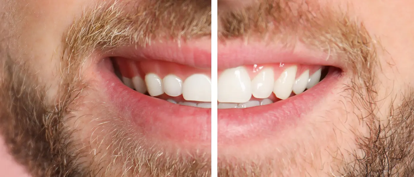
This is the most common early sign, and the most commonly dismissed. Many patients assume bleeding gums are normal, or that they're brushing "too hard." They are not normal. Healthy gums do not bleed.

Bleeding is your body's inflammatory response to bacteria that have taken up residence at or below the gumline. The gum tissue becomes swollen, fragile, and prone to bleeding with even gentle contact. This stage is called gingivitis — and it is fully reversible with proper treatment. Ignored, it progresses to periodontitis, which is not.

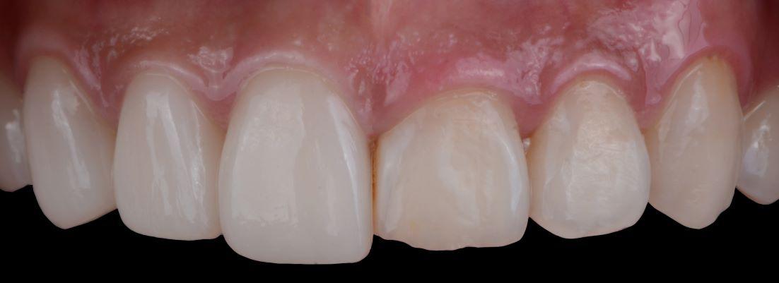
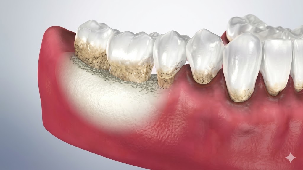
2. Gums That Look Like They're Pulling Away from Your Teeth

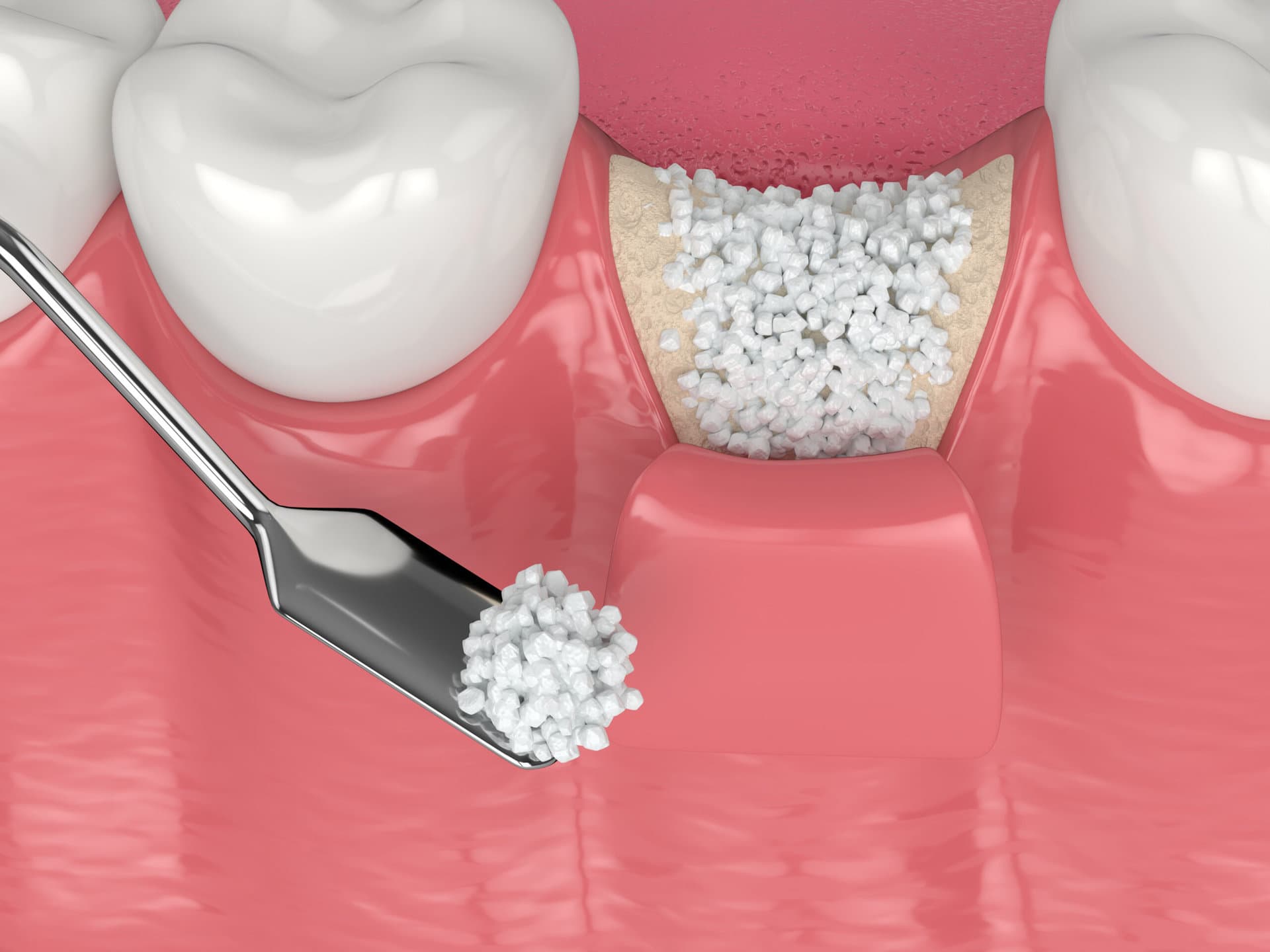
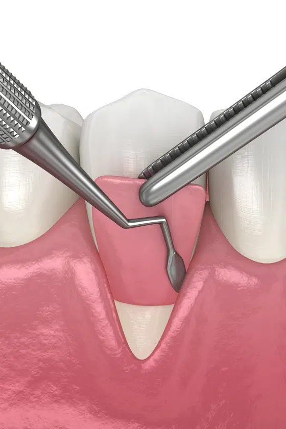
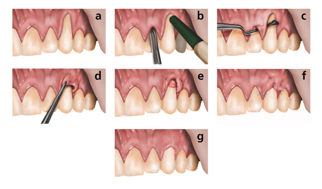
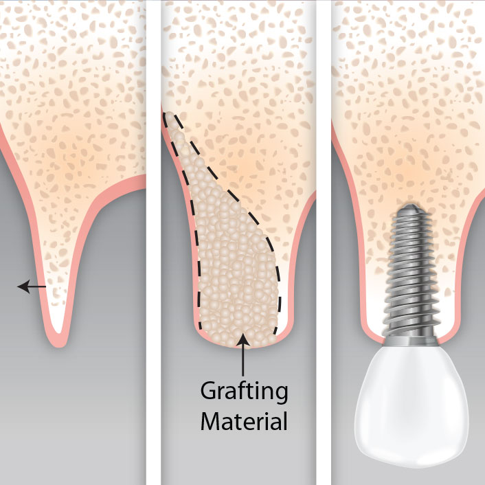
If your teeth look longer than they used to, or if you can see the yellowish root surface near the gumline, your gums may be receding. Gum recession is a hallmark sign of periodontal disease — though it can also be caused by aggressive brushing, orthodontic movement, or thin gum tissue.

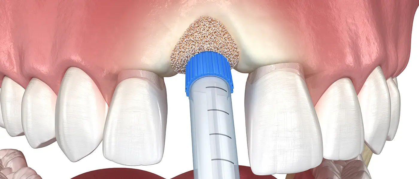
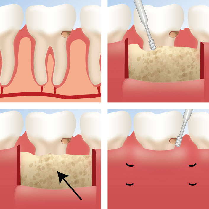
Recession exposes the tooth root, which has no enamel protection. This leads to sensitivity, especially to cold or sweet foods, and significantly increases the risk of root decay. Receded gum tissue does not grow back on its own — but it can be restored with a gum grafting procedure performed by a periodontist.

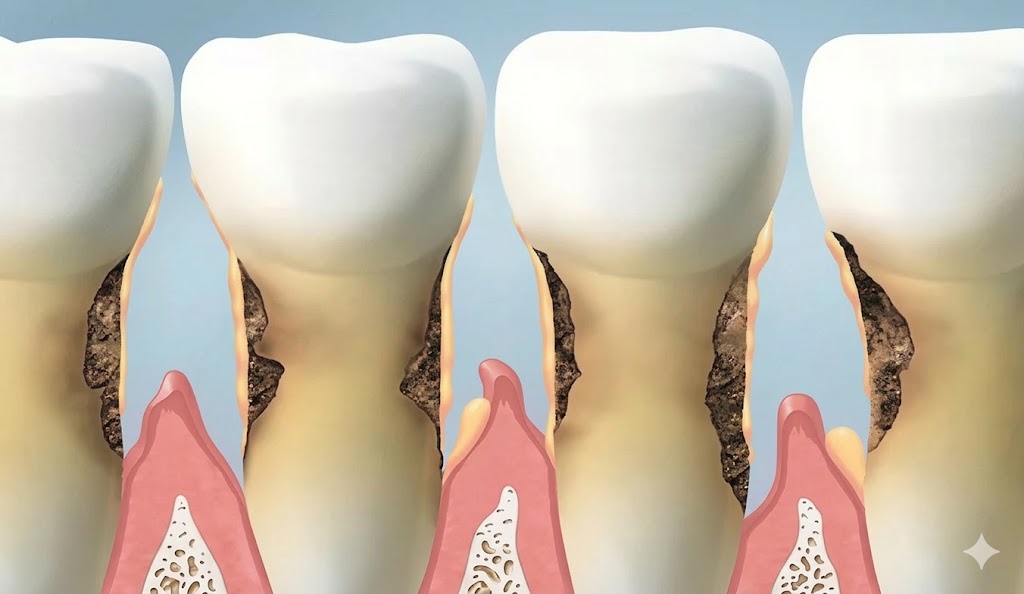
3. Persistent Bad Breath That Doesn't Respond to Brushing

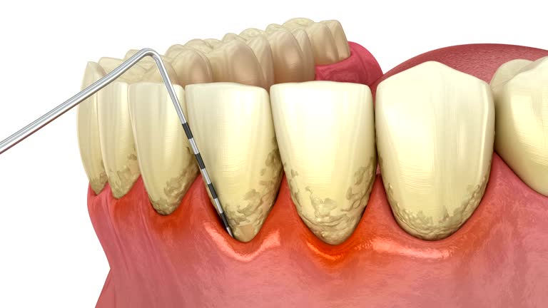
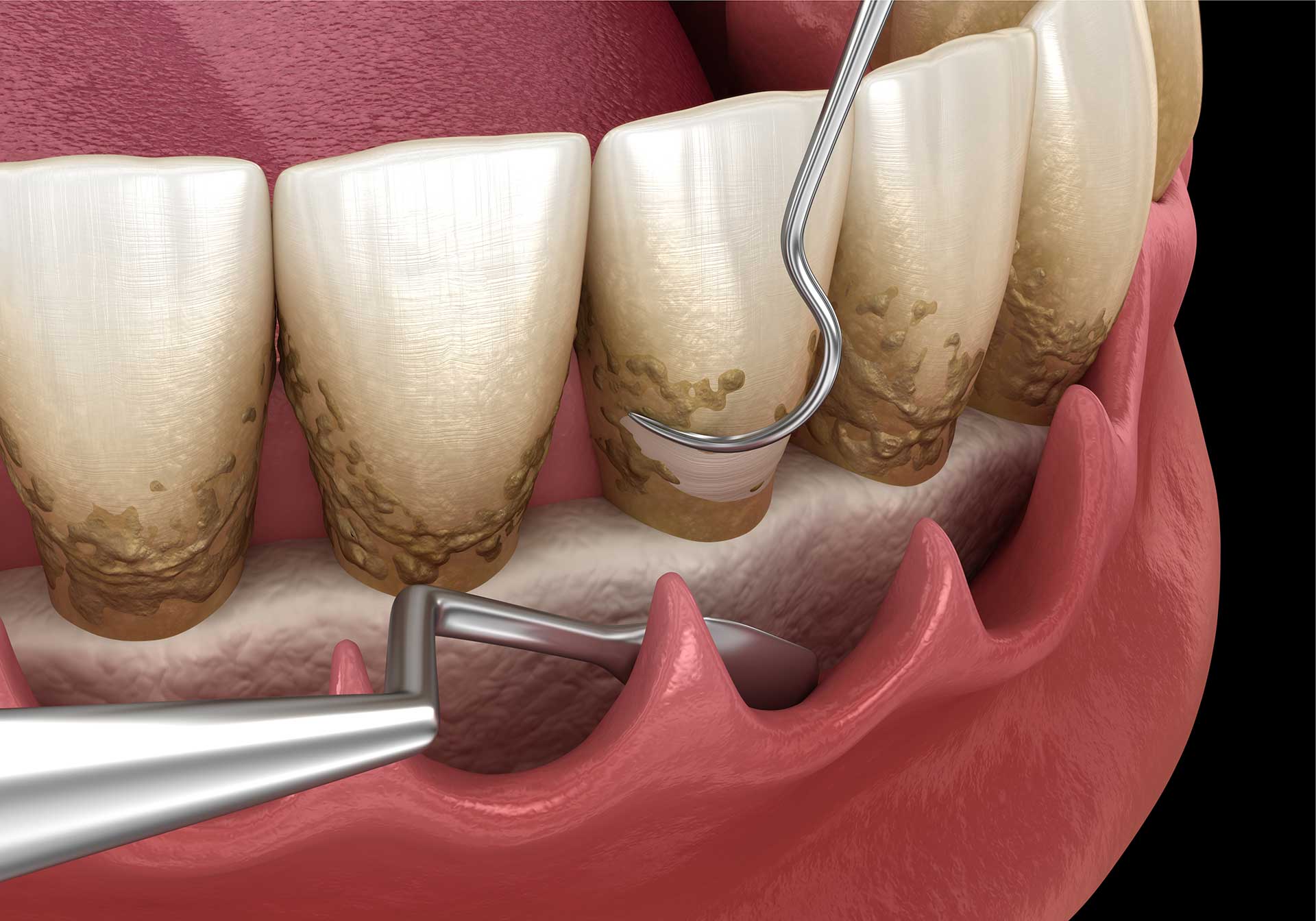
Occasional bad breath is common. Chronic halitosis that persists despite good oral hygiene is different. Bacteria living in deep periodontal pockets — the spaces between your teeth and gums that form as disease progresses — produce sulfur compounds that cause a distinctly unpleasant odor. No amount of mouthwash addresses the source.

If people in your life have mentioned bad breath, or if you notice an unpleasant taste you can't resolve, it's worth getting a periodontal evaluation.

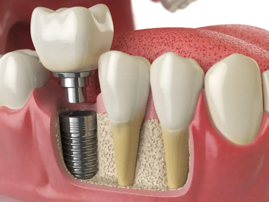
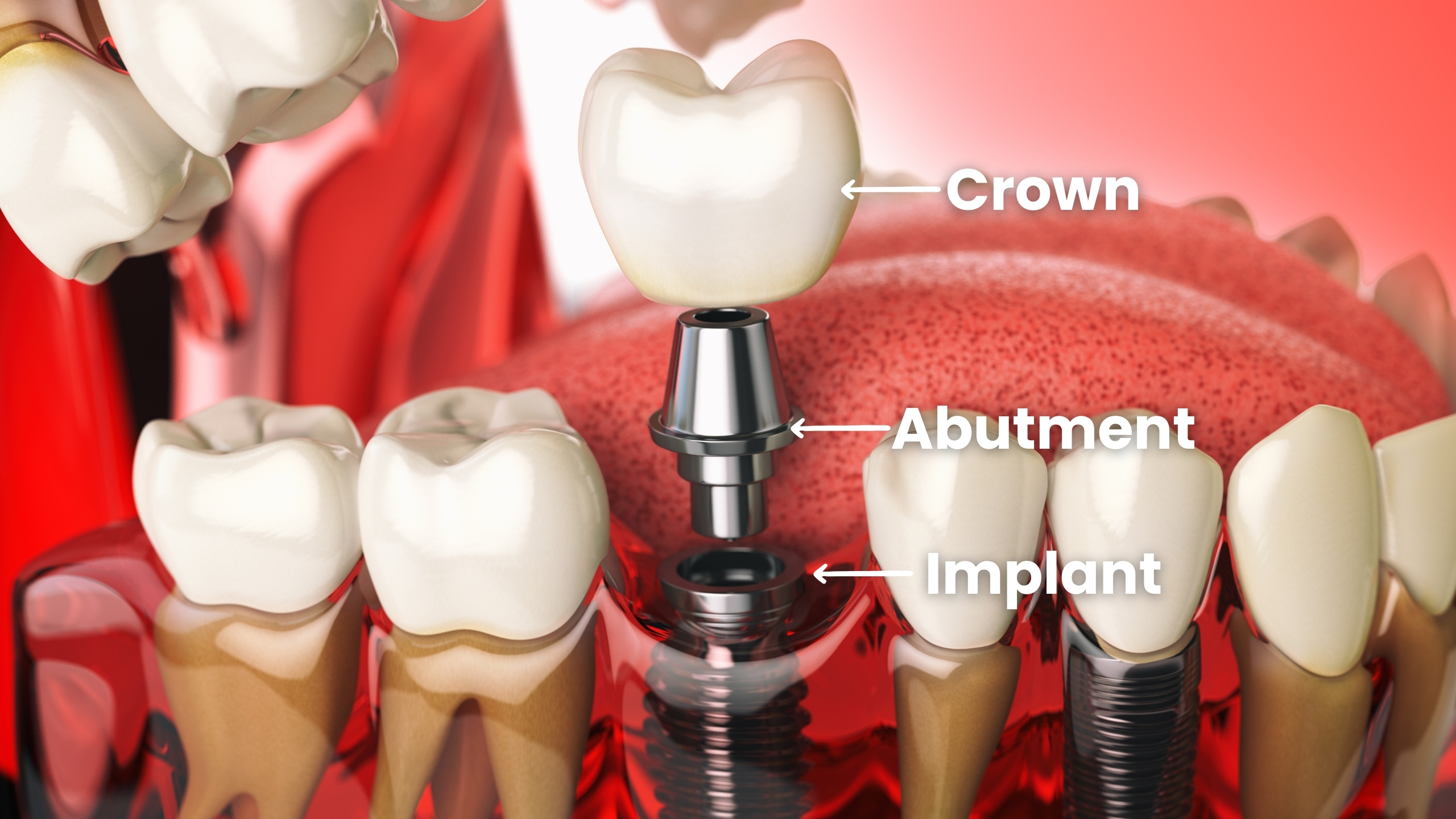
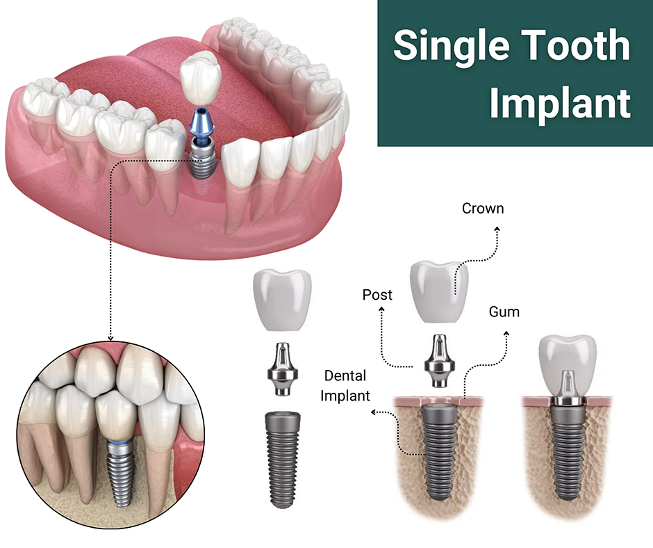
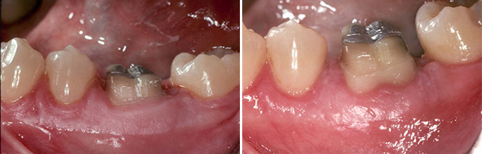
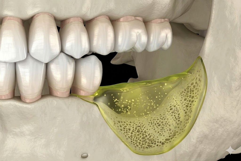
4. Teeth That Feel Loose or Have Shifted Position

Adult teeth should not feel loose. If you notice any mobility — even subtle movement when you press on a tooth — it means the supporting structures (bone and periodontal ligament) have been damaged significantly. Bone loss is the direct cause of tooth mobility in periodontal disease.

Similarly, if your bite has changed, or if teeth that were previously aligned have drifted apart or developed gaps, periodontal disease may be the cause. This degree of progression requires urgent evaluation — the goal is to preserve the remaining bone and prevent tooth loss.

5. Swollen, Red, or Tender Gum Tissue

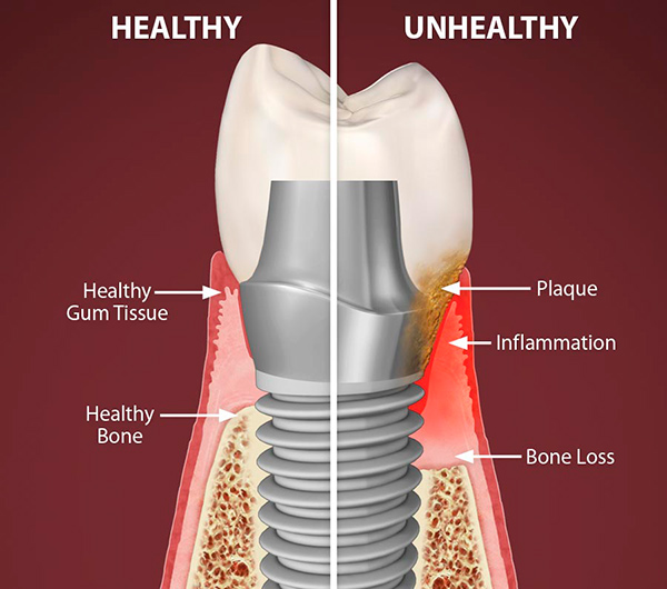
Healthy gums are firm and pale pink. Gums that are red, puffy, or tender to the touch are inflamed — a response to bacterial infection in the tissue. This can occur around individual teeth or affect the entire gum line.

Some patients notice this most during eating, when gum pressure causes discomfort. Others only notice the discoloration when looking closely in the mirror. Either way, inflamed gum tissue is not cosmetic — it reflects active infection that will progress without treatment.

What to Do If You Notice These Signs

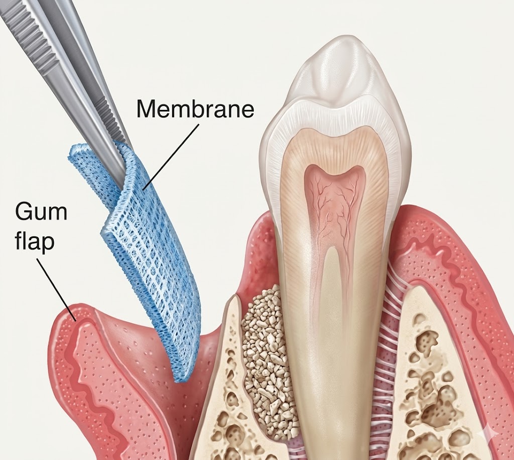
If you're experiencing one or more of the above, the right step is a comprehensive periodontal evaluation. A periodontist is a specialist who focuses exclusively on the health of the structures that support your teeth — bone, gums, and connective tissue. A thorough exam includes pocket depth measurements, bone level assessment via X-rays, and a personalized treatment plan.

Early-stage gum disease (gingivitis) can often be managed with a professional cleaning and improved home care. More advanced disease may require scaling and root planing, surgical pocket reduction, or regenerative procedures. The sooner you come in, the more options are available — and the more of your natural teeth we can preserve.

Concerned About Your Gum Health?

Schedule a comprehensive periodontal evaluation with Dr. Ramin Foroughi in Oxnard, CA. Most patients are surprised at how painless and straightforward the process is.

HomeBlog › Implants vs. Dentures

Dental Implants vs. Dentures: Which Is Right for You?

When patients lose multiple teeth — or face losing them — they're often presented with two options: dentures or dental implants. Both replace missing teeth. But they work very differently, and their long-term outcomes are not equivalent. Here's an honest comparison from a periodontist who places dental implants every day.

The Fundamental Difference

The most important distinction between implants and dentures is what they do — or don't do — for your jawbone.

Your jawbone is maintained by the stimulation of tooth roots pressing against it during chewing. When a tooth is lost, the bone beneath it no longer receives that stimulation. The body interprets this as a signal to resorb the bone — and it begins doing so within weeks of extraction. Over the first year after tooth loss, up to 25% of bone volume can be lost. This accelerates over time.

Dentures sit on top of the gum and bone — they do not replace the tooth root and provide no stimulation. The bone loss continues underneath them, which is why dentures that fit well initially often become loose over the years. Dental implants, by contrast, are placed directly into the jawbone and fuse with it through a process called osseointegration. They function as tooth roots, preventing bone loss entirely.

Comfort and Function

Dentures are removable prosthetics that rest on the gums. Lower dentures in particular are notoriously unstable, because the lower arch has less surface area to grip. Many patients find that lower dentures rock, shift, and limit their diet significantly. Adhesives help temporarily but don't solve the underlying instability. As bone continues to resorb, fit deteriorates further.

Implants are fixed in the jaw and function like natural teeth. Patients bite, chew, and speak without thinking about their teeth — because the implants behave exactly as roots do. There is no movement, no adhesive, no dietary restriction, and no removal at night. Most patients report that implants are indistinguishable from their natural teeth in everyday function.

Cost: The Real Comparison

Dentures have a lower upfront cost, which makes them attractive. But cost comparisons should account for the full picture:

  • Dentures require relining as bone resorbs and the fit changes — typically every few years
  • Poorly fitting dentures accelerate bone loss, potentially requiring more complex (and more expensive) bone grafting if implants are pursued later
  • The longer a patient waits to pursue implants after tooth loss, the more bone is gone — meaning more extensive (and more costly) pre-implant bone grafting may be needed
  • Implants, maintained well, last decades. The per-year cost over a lifetime is often lower than repeatedly replacing dentures

We understand that implants are a significant investment upfront, and we work with several financing partners to make treatment accessible. But patients should make this decision with full information — not just the initial sticker price.

Implant-Supported Dentures: The Middle Ground

For patients who want the stability of implants but at a lower cost than replacing every tooth individually, implant-supported overdentures offer a compelling option. Two to four implants are placed in the jaw, and the denture "snaps on" to them. The result is dramatically better stability than a conventional denture — and the implants prevent the bone loss that makes conventional dentures problematic over time.

For patients who are missing all their teeth, All-On-4 or All-On-6 full-arch implants provide a completely fixed bridge anchored to four or six implants. The result is a full set of teeth that do not come out — permanent, stable, and bone-preserving.

Who Is a Candidate for Implants?

Most healthy adults who have lost teeth are candidates for dental implants. The key requirements are:

  • Sufficient jawbone volume to anchor the implant (if not, bone grafting can often create it)
  • Healthy gums free of active infection
  • No uncontrolled systemic conditions that would impair healing

Even patients who have been told they "don't have enough bone" for implants are often candidates after a bone grafting procedure. A 3D CBCT scan gives us the precise information needed to evaluate what's possible for each patient specifically.

Our Recommendation

For most patients, dental implants are the superior long-term solution. They preserve bone, function like natural teeth, and eliminate the ongoing complications of removable dentures. The question isn't really "implants or dentures" — it's "what type of implant solution is right for your situation and budget?" We're happy to walk through all the options at a consultation.

Ready to Explore Your Options?

Schedule a dental implant consultation with Dr. Ramin Foroughi in Oxnard, CA. We'll review your bone levels, explain all options, and give you a clear picture of what's possible.

HomeBlog › What Is a Sinus Lift?

What Is a Sinus Lift — and Do You Need One for Dental Implants?

If you've been told you need a sinus lift before getting dental implants in your upper back jaw, you might be wondering what that means — and whether it's really necessary. This is one of the most common questions we get from patients evaluating their implant options, so let's walk through it clearly.

Why the Upper Back Jaw Is Different

Your maxillary sinuses are large air-filled cavities that sit just above your upper back teeth (molars and premolars). In most people, the floor of the sinus sits fairly close to the roots of those teeth.

When an upper back tooth is lost, two things happen simultaneously: the jawbone beneath the extraction site begins to resorb (shrink), and the sinus floor can gradually expand downward into the space vacated by the lost root. The result, over time, is a situation where there may not be enough vertical bone height between the gum surface and the sinus floor to safely place a dental implant — which needs at least 10–12mm of bone to be anchored securely.

A sinus lift solves this problem by elevating the sinus membrane and placing bone graft material in the created space, building the bone height needed to support an implant.

What the Procedure Involves

There are two techniques, chosen based on how much additional bone height is needed:

Internal (Crestal) Sinus Lift

When only a small amount of bone needs to be added (typically less than 4–5mm), an internal sinus lift can be performed through the same access point used for the implant. A special instrument gently elevates the sinus membrane through the implant site, and graft material is packed into the created space. In many cases, the implant can be placed at the same appointment.

Lateral (Window) Sinus Lift

When more significant bone volume is needed, the procedure is performed through a small opening ("window") made in the side of the jaw. The sinus membrane is carefully lifted away from the bone, graft material is placed in the space, and the window is covered with a protective membrane. The site is allowed to heal for approximately 6–9 months before implant placement.

Both procedures are performed under local anesthesia in our office, with sedation available for anxious patients. Most people are surprised at how manageable the experience is.

Recovery: What to Expect

The first 2–3 days after a lateral sinus lift are typically the most uncomfortable. Patients can expect:

  • Mild to moderate swelling of the cheek and under the eye
  • Some bruising, particularly on the cheek
  • Slight congestion or blood-tinged nasal discharge for a few days
  • Soreness well-managed with prescribed or over-the-counter pain medication

Most patients return to office work within 2–3 days. Heavy exercise and activities that involve straining or blowing the nose (which could disturb the graft) should be avoided for 2–3 weeks.

An internal sinus lift involves less recovery — most patients feel back to normal within a day or two.

Is a Sinus Lift Right for You?

Not every patient with upper back tooth loss needs a sinus lift. It depends on how much bone remains, where the sinus floor sits, and which implant position is needed. A 3D CBCT scan gives us precise measurements and allows us to plan exactly what's required.

Some patients are surprised to learn that a sinus lift they were dreading turns out to be a simple internal procedure done at the time of implant placement — not a separate surgery at all. Others do require the more involved lateral approach, but find recovery completely manageable with proper preparation and aftercare guidance.

The bottom line: a sinus lift is a well-established, highly predictable procedure that opens the door to implants for patients who would otherwise not be candidates. If you've been told you need one, it's not a red flag — it's a solution.

Find Out If You Need a Sinus Lift

A 3D CBCT scan and evaluation with Dr. Foroughi will tell you exactly what's needed — and what's possible — for your upper back teeth. Serving Oxnard and Ventura County.

HomeBlog › Why Gum Recession Happens

Why Gum Recession Happens — and How to Stop It

Gum recession is one of the most common reasons patients are referred to a periodontist. You might notice it as sensitivity to cold, a tooth that looks longer than it used to, or an exposed yellowish root near the gumline. Left untreated, recession leads to root damage, decay in areas that were never meant to be exposed, and eventually — if the supporting tissue is lost entirely — tooth loss.

The encouraging news is that gum recession is very treatable. The key is understanding why it's happening, because the cause shapes the treatment.

The Most Common Causes of Gum Recession

1. Periodontal Disease

Bacterial infection in the tissue around the teeth is the leading cause of gum recession in adults. As the bacteria inflame and destroy the gum tissue and underlying bone, the gumline migrates downward. This type of recession typically affects multiple teeth and is accompanied by other signs of gum disease — bleeding, swelling, bone loss, and pocket formation.

2. Aggressive or Incorrect Brushing

Brushing too hard — or using a stiff-bristled toothbrush — physically abrades the gum tissue over years. This is called toothbrush abrasion, and it tends to affect isolated teeth (often on the side of the dominant hand) with a characteristic notched or worn pattern at the gumline. The gum tissue is worn away mechanically, not by bacteria.

3. Thin Gum Tissue (Thin Biotype)

Some people are born with naturally thin, delicate gum tissue — a characteristic called a "thin periodontal biotype." This tissue is more prone to recession from any mechanical or inflammatory trigger. It's not the patient's fault; it's anatomy. But it does mean that proactive treatment (gum grafting to thicken the tissue) is often appropriate before recession becomes more severe.

4. Misaligned Teeth or Bite Issues

Teeth that are positioned outside the normal arch — especially teeth that tip outward — often have very thin or absent bone on their outer surface. This makes the overlying gum tissue vulnerable to recession. Orthodontic movement can sometimes worsen recession in these situations if not carefully planned.

5. Tooth Grinding (Bruxism)

Chronic grinding generates lateral forces on the teeth that can damage the periodontal attachment over time, contributing to recession in some patients.

What Happens If You Leave It Untreated

Gum tissue that has receded does not grow back on its own. Once root surface is exposed, it stays exposed — and the recession typically progresses. Exposed roots are:

  • Vulnerable to decay (root cementum is softer than enamel)
  • Sensitive to temperature, sweets, and touch
  • More prone to physical wear from brushing
  • Aesthetically visible when smiling

Beyond the tooth itself, significant recession usually indicates loss of the underlying supporting bone — which is the real long-term threat to keeping the tooth.

How It's Treated: Gum Grafting

The most effective treatment for gum recession is a connective tissue graft (CTG) — the gold standard procedure for restoring receded gum tissue. In this procedure, a small amount of tissue is taken from the roof of the mouth and placed precisely over the exposed root surface, extending the gumline back to where it belongs.

The graft serves multiple purposes: it covers and protects the exposed root, thickens the gum tissue to resist future recession, and restores the natural contour and appearance of the gumline. In appropriate cases, complete root coverage is achievable — meaning the recession is effectively eliminated.

The procedure is performed under local anesthesia and takes approximately one hour. Recovery is manageable for most patients, with soft diet restrictions for about two weeks while the graft heals.

The Right Time to Treat Recession

Earlier is always better. Small areas of recession are easier to treat, require less donor tissue, and have higher rates of complete root coverage. If you notice that a tooth looks longer, feels sensitive at the gumline, or if you can see the root surface, that's the time to get an evaluation — not after waiting another year to see if it gets worse.

If the recession is due to periodontal disease, the underlying infection must be treated first — before grafting. If it's due to brushing technique, that habit must be corrected alongside the surgical correction.

Worried About Receding Gums?

Dr. Ramin Foroughi specializes in gum grafting and periodontal disease treatment in Oxnard, CA. An evaluation will tell you exactly where you stand and what your options are.